Abdominal Blood Vessels Labeled : Perineum Stab Yes Really Fightwrite / Abdominal vessels are typically imaged in several planes (fig.. Abdominal vessels are typically imaged in several planes (fig. The videos are done by dr. Learn everything about the blood vessels of the abdominal wall with our article, quizzes, and labeled diagrams. Before lab, read through the procedure and draw diagrams of the blood vessels you will be finding and label these. The aorta is the large artery leaving the heart.
3 the superficial vessels include the superficial epigastric and the superficial circumflex iliac vessels. Advertising on our site helps support our mission. These vessels usually include the abdominal aorta and/or inferior vena cava (ivc). We will include an analysis of the normal doppler waveforms of the abdominal vessels. We will include an analysis of the normal doppler waveforms of the abdominal vessels.
Abdominal Aortic Aneurysm Doctor Stock from m.psecn.photoshelter.com Teachme anatomy part of the teachme series the medical information on this site is provided as an information resource only, and is not to be used or relied on for any diagnostic or treatment purposes. Aorta (aorta) the aorta is the first segment of the systemic arterial circulation, originating directly from the left ventricle of the heart.it is the largest artery in the body consisting of three parts that each has its special characteristics, most notably in their direction and orientation. The common iliac arteries and veins. Katy wallis at state college of florida As the abdomen and pelvis contain the majority of internal organs, these regions need to be supplied by an extensive network of arteries and veins. Label the biliary passages and associated structures using the hints provided. Label the intestinal structures using the hints provided. Veins are vessels that return.
Of course, recognition of the normal vascular anatomy is essential for the investigation of any abdominal vascular problem.
We will include an analysis of the normal doppler waveforms of the abdominal vessels. Neurovasculature of the abdominal wall explore study unit superior epigastric artery: Aorta (aorta) the aorta is the first segment of the systemic arterial circulation, originating directly from the left ventricle of the heart.it is the largest artery in the body consisting of three parts that each has its special characteristics, most notably in their direction and orientation. Label the intestinal structures using the hints provided. Advertising on our site helps support our mission. These vessels are branches of the femoral artery and vein. By switching the orientation of the probe, it is possible to measure the diameter of the vessels, the origin of vessels, and their angles as they take off from the abdominal aorta. • they are also common in abdominal organs, the heart, and the brain dr. Doppler studies of the abdominal vessels demand an understanding of normal and abnormal blood flow patterns. 3 the superficial vessels include the superficial epigastric and the superficial circumflex iliac vessels. Abdominal blood vessels labeled / a p 2 lab test 2 flashcards quizlet : Want to learn more about it? Blood vessels of the abdomen and pelvis.
In human anatomy, inferior epigastric artery refers to the artery that arises from the external iliac artery.it anastomoses with the superior epigastric artery.along its course, it is accompanied by a similarly named vein, the inferior epigastric vein.these epigastric vessels form the lateral border of hesselbach's triangle, which outlines the area through which direct inguinal hernias protrude. Physiology and anatomy of blood vessels prepared by dr. Veins are vessels that return. Abdominal vessels are typically imaged in several planes (fig. The videos are done by dr.
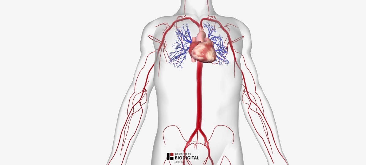
Arteries Of The Body Picture Anatomy Definition More from human.biodigital.com • they are also common in abdominal organs, the heart, and the brain dr. Label the biliary passages and associated structures using the hints provided. Blood vessels of the abdomen and pelvis. Teachme anatomy part of the teachme series the medical information on this site is provided as an information resource only, and is not to be used or relied on for any diagnostic or treatment purposes. That being said, all arterial blood delivered to this region comes via branches of the abdominal aorta, and all venous blood eventually finds its way back to. Want to learn more about it? Of course, recognition of the normal vascular anatomy is essential for the investigation of any abdominal vascular problem. By switching the orientation of the probe, it is possible to measure the diameter of the vessels, the origin of vessels, and their angles as they take off from the abdominal aorta.
Label the intestinal structures using the hints provided.
Katy wallis at state college of florida Label the intestinal structures using the hints provided. Of course, recognition of the normal vascular anatomy is essential for the investigation of any abdominal vascular problem. The superior vena cava is the large vein that brings blood from the head and arms to the heart, and the inferior vena cava brings blood from the abdomen and legs into the heart. Practice identifying the blood vessels on the photographs here and in your fetal pig photoalbum online. 3 the superficial vessels include the superficial epigastric and the superficial circumflex iliac vessels. Dissection of the blood vessels posterior to the diaphragm procedure: That being said, all arterial blood delivered to this region comes via branches of the abdominal aorta, and all venous blood eventually finds its way back to. As the abdomen and pelvis contain the majority of internal organs, these regions need to be supplied by an extensive network of arteries and veins. Want to learn more about it? Label the abdominal blood vessels using the hints provided. Structure of blood vessel walls. Before lab, read through the procedure and draw diagrams of the blood vessels you will be finding and label these.
Learn everything about the blood vessels of the abdominal wall with our article, quizzes, and labeled diagrams. The identification of abdominal vessels using ultrasound is based on knowledge of their normal location, appearance and relationship to specific organs. Label the biliary passages and associated structures using the hints provided. The common iliac arteries and veins. It supplies blood to the internal and external carotid arteries.
Arteries Of Posterior Abdominal Wall Blood Supply Of The Abdomen from netterimages.com Teachme anatomy part of the teachme series the medical information on this site is provided as an information resource only, and is not to be used or relied on for any diagnostic or treatment purposes. Label the intestinal structures using the hints provided. The videos are done by dr. Abdominal blood vessels labeled / a p 2 lab test 2 flashcards quizlet : • they are also common in abdominal organs, the heart, and the brain dr. Anatomy of blood vessels of abdomen pelvic cavities. Neurovasculature of the abdominal wall explore study unit superior epigastric artery: By switching the orientation of the probe, it is possible to measure the diameter of the vessels, the origin of vessels, and their angles as they take off from the abdominal aorta.
That being said, all arterial blood delivered to this region comes via branches of the abdominal aorta, and all venous blood eventually finds its way back to.
Label the biliary passages and associated structures using the hints provided. Blood vessels the major vessels in the anterior abdominal wall can be divided into deep and superficial vessels (fig. Practice identifying the blood vessels on the photographs here and in your fetal pig photoalbum online. • they are also common in abdominal organs, the heart, and the brain dr. Label the biliary passages and associated structures using the hints provided. By switching the orientation of the probe, it is possible to measure the diameter of the vessels, the origin of vessels, and their angles as they take off from the abdominal aorta. The aorta is the large artery leaving the heart. They ultimately bifurcate into a (right and left) external and internal carotid artery at the superior border of the laryngeal thyroid cartilage. Blood vessels of the abdomen and pelvis. Before lab, read through the procedure and draw diagrams of the blood vessels you will be finding and label these. Anatomy of blood vessels of abdomen pelvic cavities. Dissection of the blood vessels posterior to the diaphragm procedure: Label the abdominal blood vessels using the hints provided.
The videos are done by dr blood vessels labeled. These vessels usually include the abdominal aorta and/or inferior vena cava (ivc).


0 Komentar